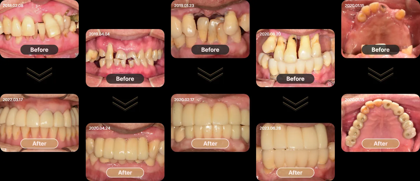
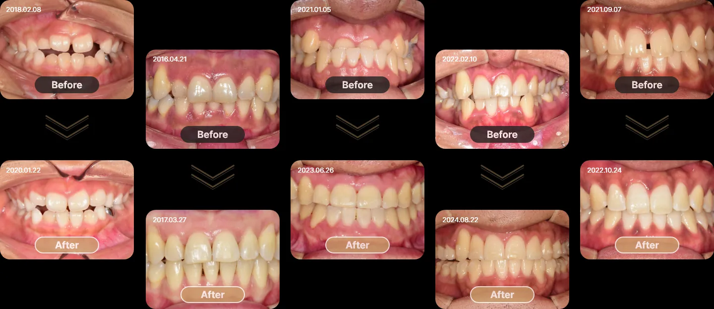
Cases are published with the patients’ consent. Treatment plans, duration and results differ for every patient, and every treatment carries risks such as sensitivity, swelling or the need for later adjustment. Only an examination can show what is possible for you.
Implants and full-mouth restoration
Missing and failing teeth replaced with implant-supported crowns and bridges. Each pair shows the same patient before and after treatment. About this treatment →
Invisalign
Crowding, spacing and protruding teeth treated with Invisalign clear aligners. Each pair shows the same patient before and after treatment. About this treatment →
Implant cases
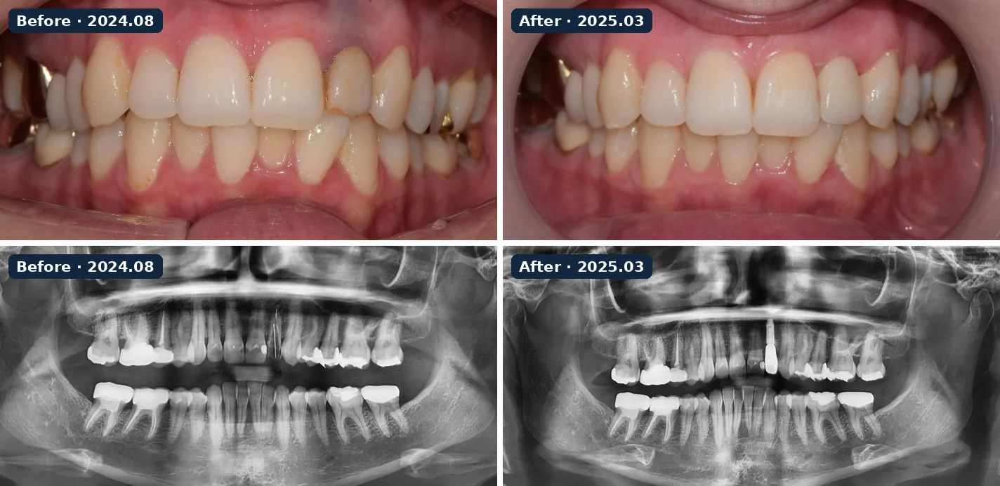
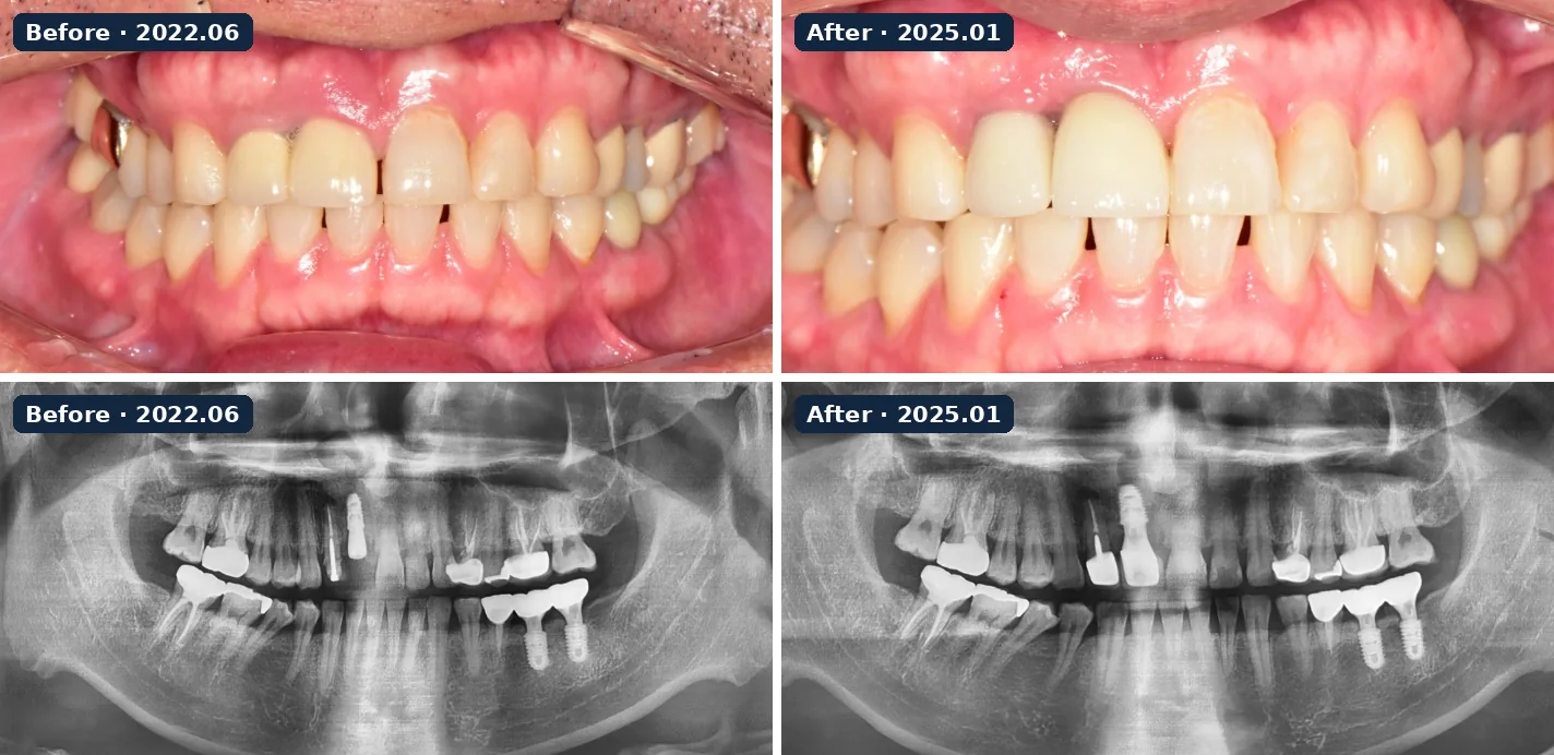
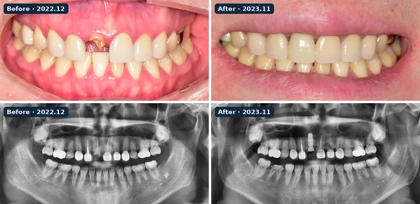
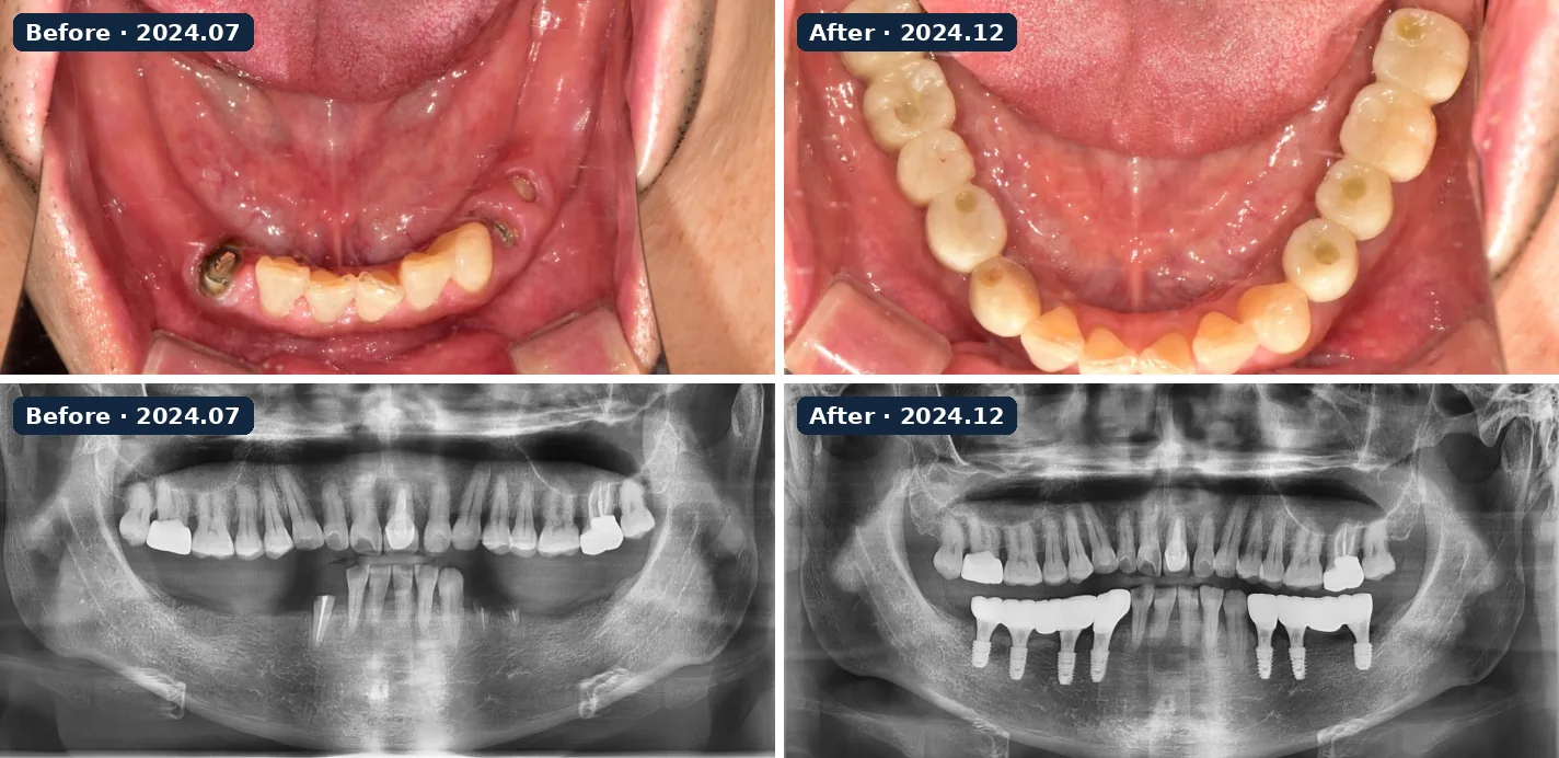
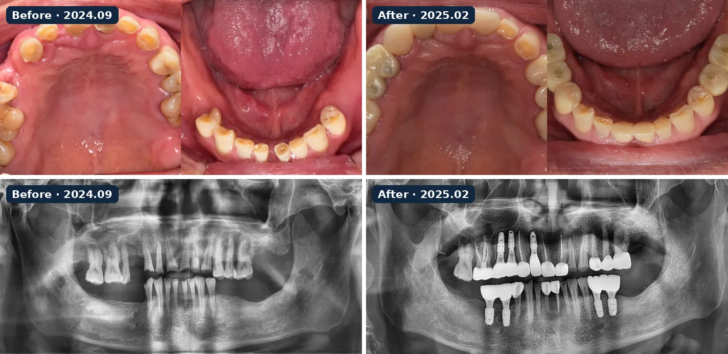
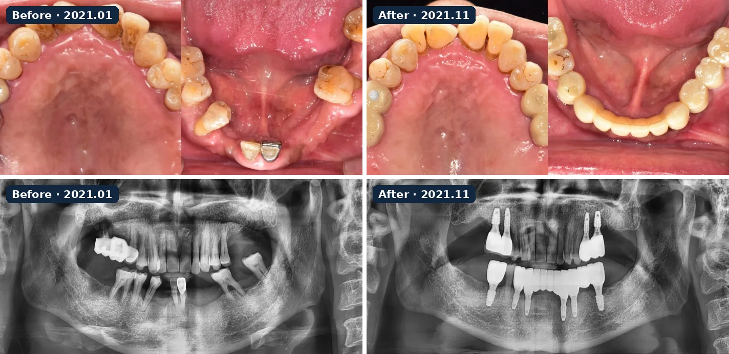
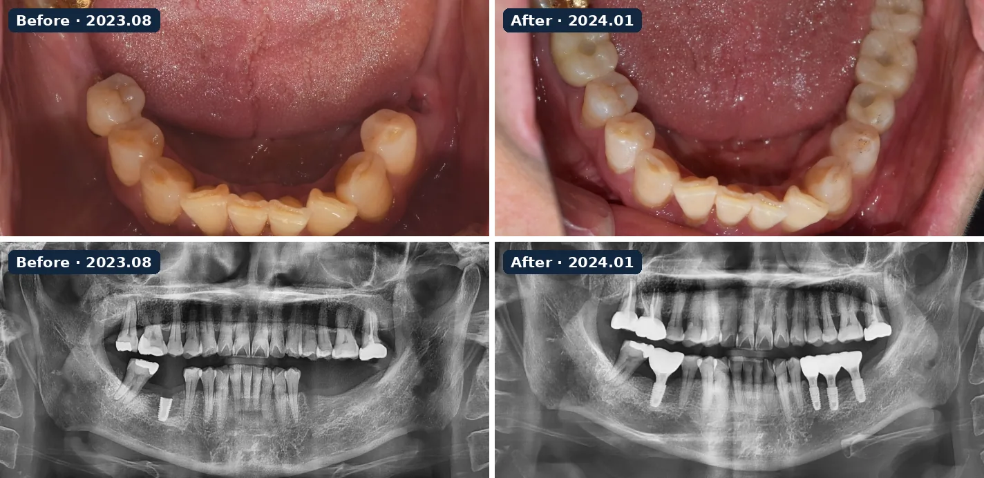
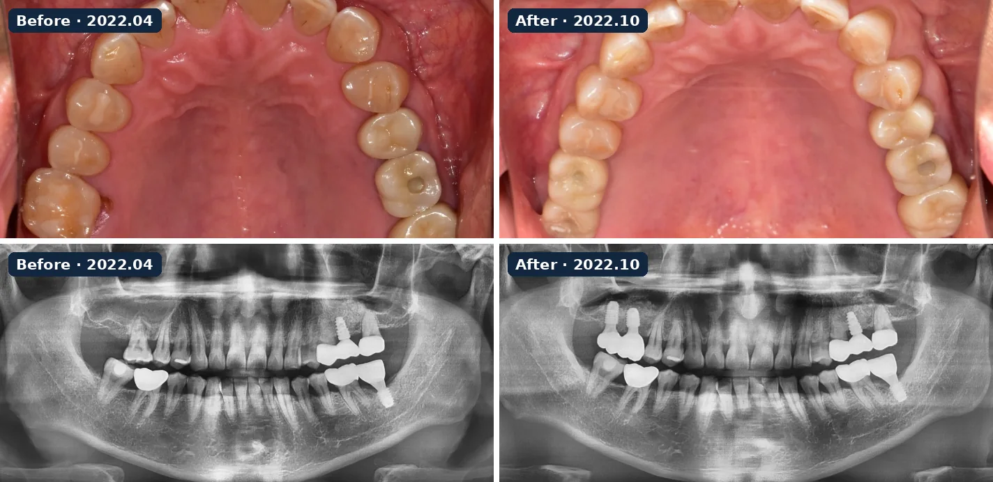
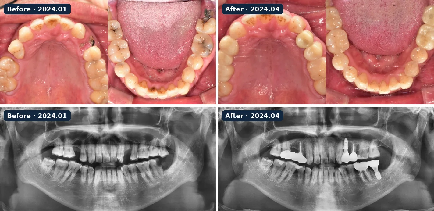
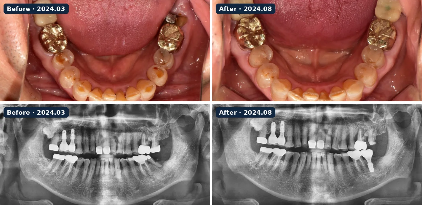
Ten implant cases from the clinic’s records, each showing the same patient before and after treatment with intraoral photos and panoramic X-rays. Published with the patients’ consent; before and after images were taken under the same conditions. Results differ for every patient. If aftercare is neglected, implants can develop bleeding or peri-implantitis, so good oral hygiene and regular check-ups matter. About dental implants →
Implant case 1 · 2024.08 → 2025.03 · intraoral photos and panoramic X-raysImplant case 2 · 2022.06 → 2025.01 · intraoral photos and panoramic X-raysImplant case 3 · 2022.12 → 2023.11 · intraoral photos and panoramic X-raysImplant case 4 · 2024.07 → 2024.12 · intraoral photos and panoramic X-raysImplant case 5 · 2024.09 → 2025.02 · intraoral photos and panoramic X-raysImplant case 6 · 2021.01 → 2021.11 · intraoral photos and panoramic X-raysImplant case 7 · 2023.08 → 2024.01 · intraoral photos and panoramic X-raysImplant case 8 · 2022.04 → 2022.10 · intraoral photos and panoramic X-raysImplant case 9 · 2024.01 → 2024.04 · intraoral photos and panoramic X-raysImplant case 10 · 2024.03 → 2024.08 · intraoral photos and panoramic X-rays
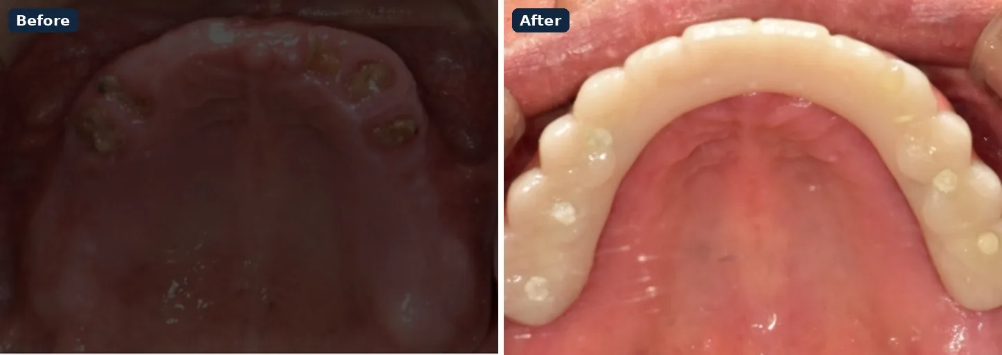
Full-arch case
Patient without teeth for several years; six implants placed in each jaw with a digitally planned full-arch prosthesis.
Laminate veneers
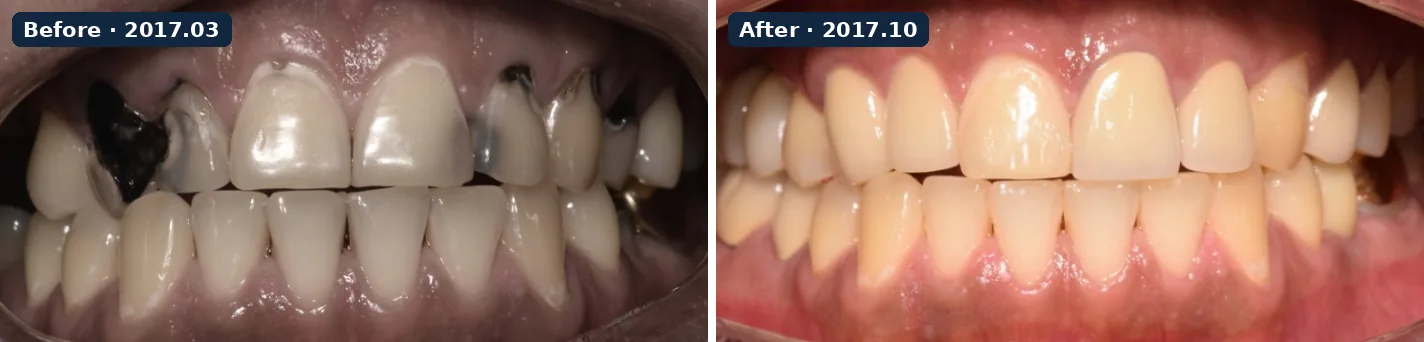
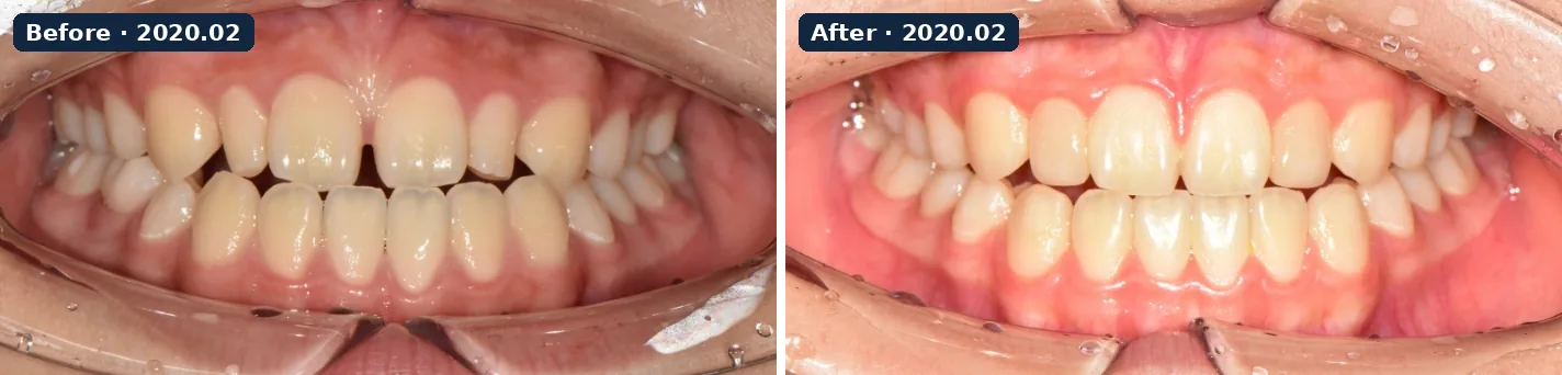
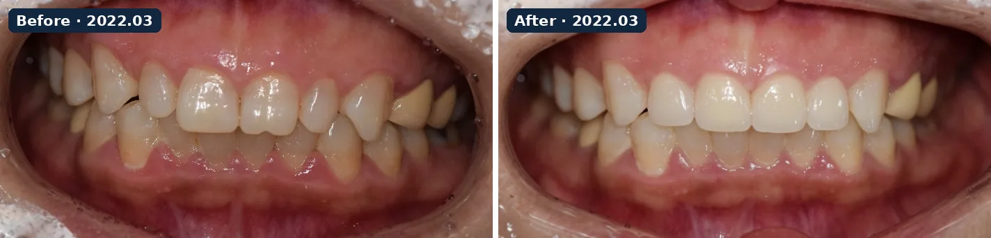
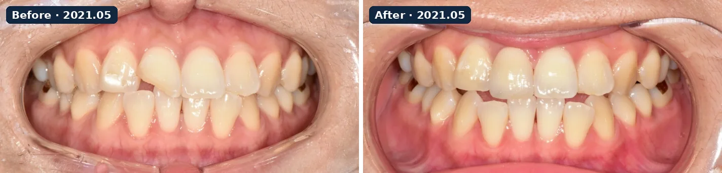
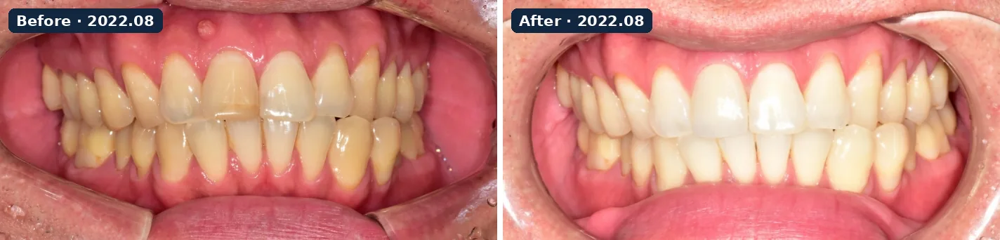
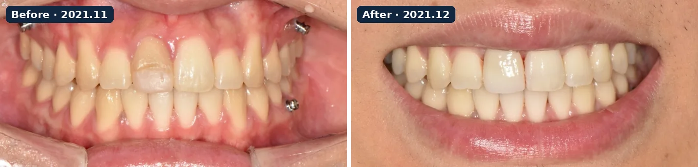
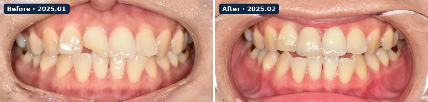
Seven veneer cases from the clinic’s records, each showing the same patient before and after treatment. Published with the patients’ consent; before and after images were taken under the same conditions. Results differ for every patient. Veneers can cause tooth sensitivity or chipping of the restoration, so accurate diagnosis and aftercare matter. About veneers →
Laminate veneer case 1 · 2017.03 → 2017.10 · intraoral photos · veneers combined with an implantLaminate veneer case 2 · 2020.02 → 2020.02 · intraoral photosLaminate veneer case 3 · 2022.03 → 2022.03 · intraoral photosLaminate veneer case 4 · 2021.05 → 2021.05 · intraoral photosLaminate veneer case 5 · 2022.08 → 2022.08 · intraoral photosLaminate veneer case 6 · 2021.11 → 2021.12 · intraoral photosLaminate veneer case 7 · 2025.01 → 2025.02 · intraoral photos
Orthodontics (Invisalign page cases)
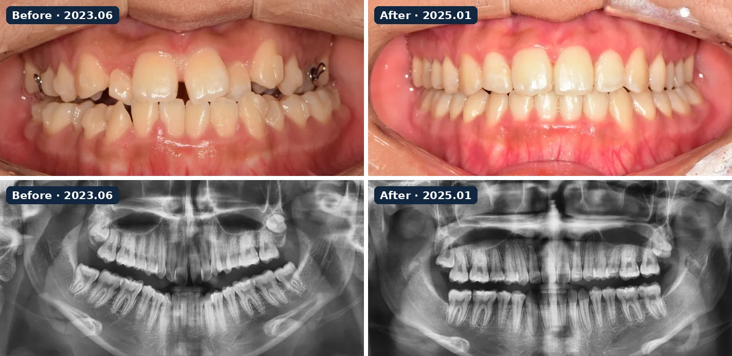
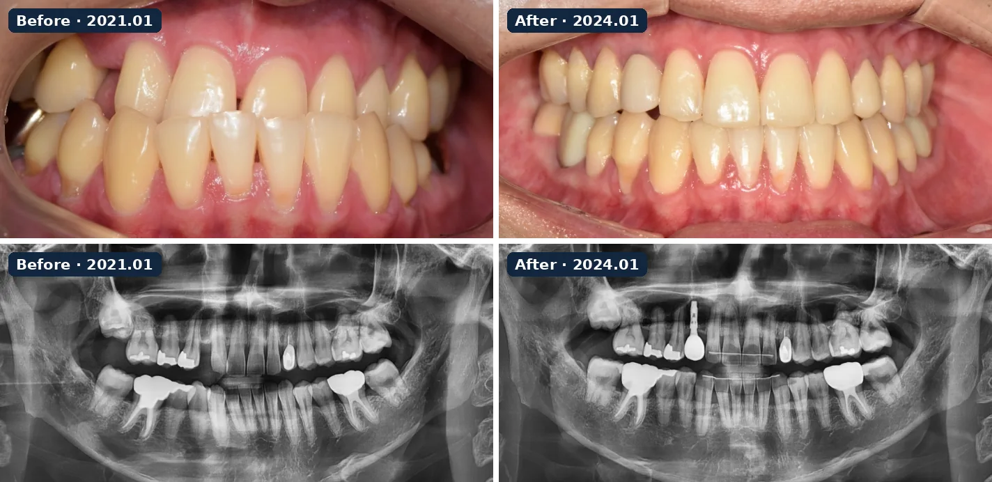
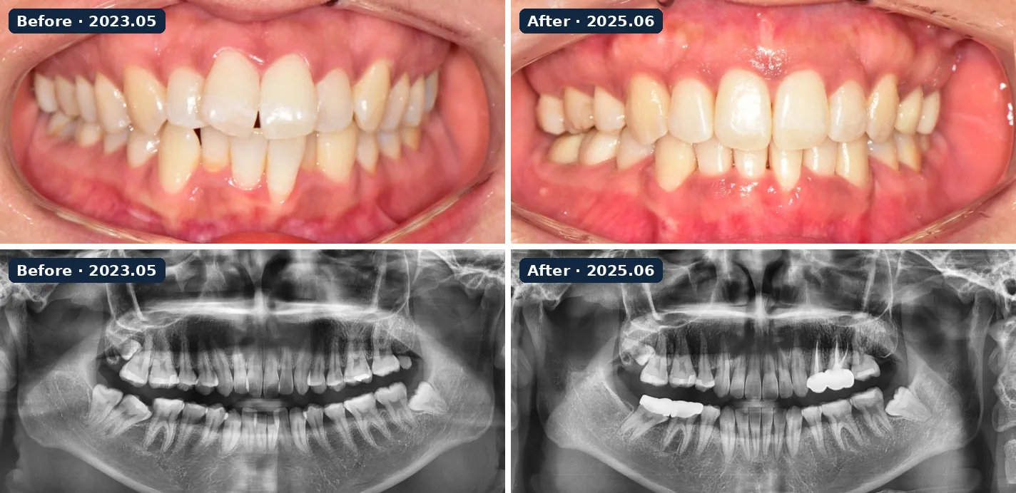
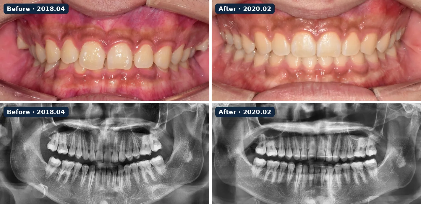
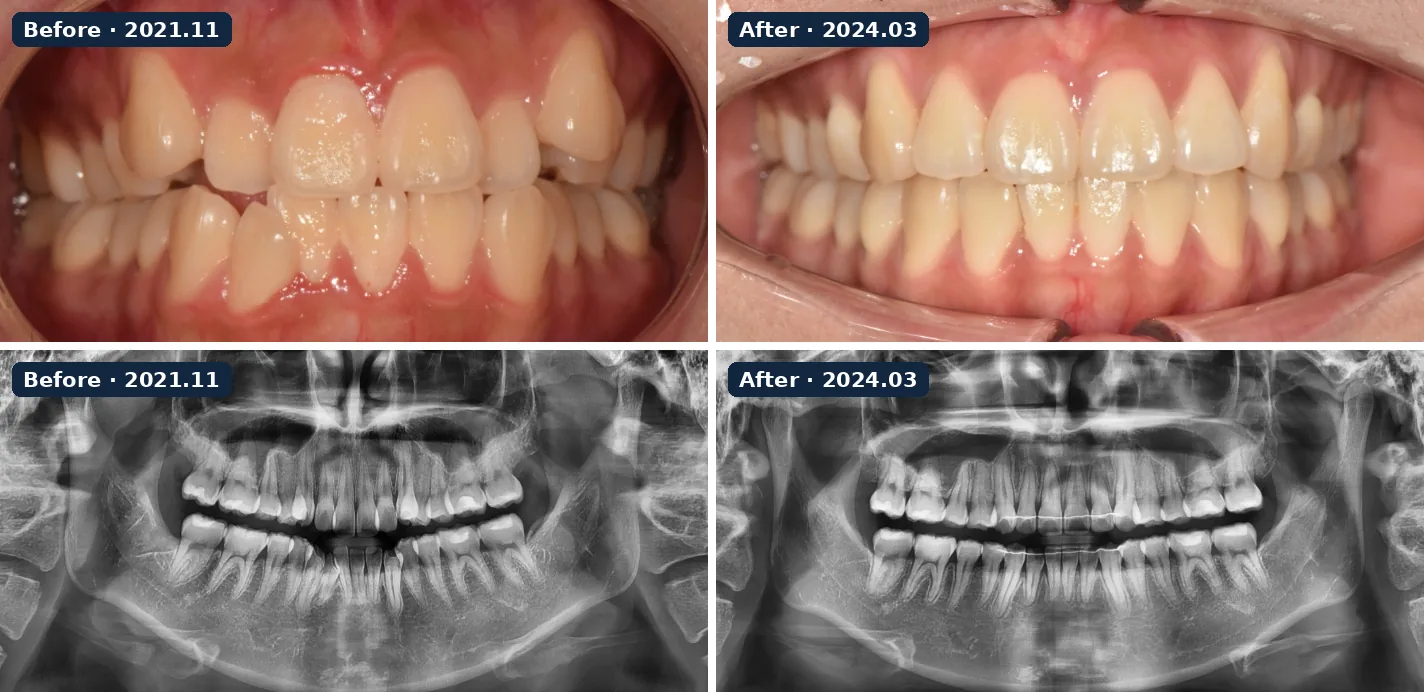
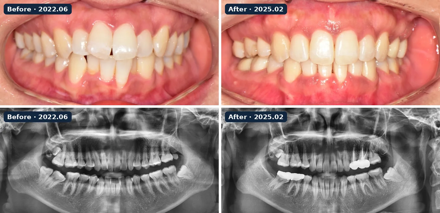
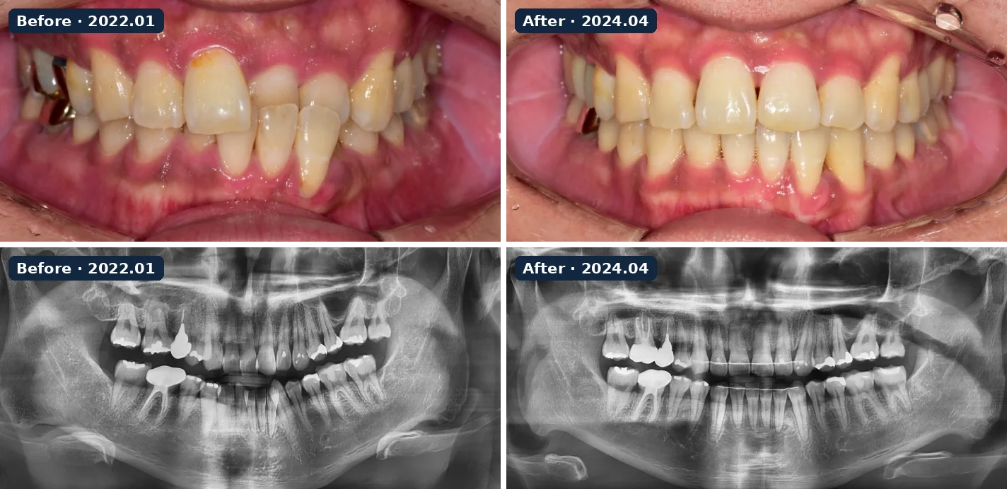
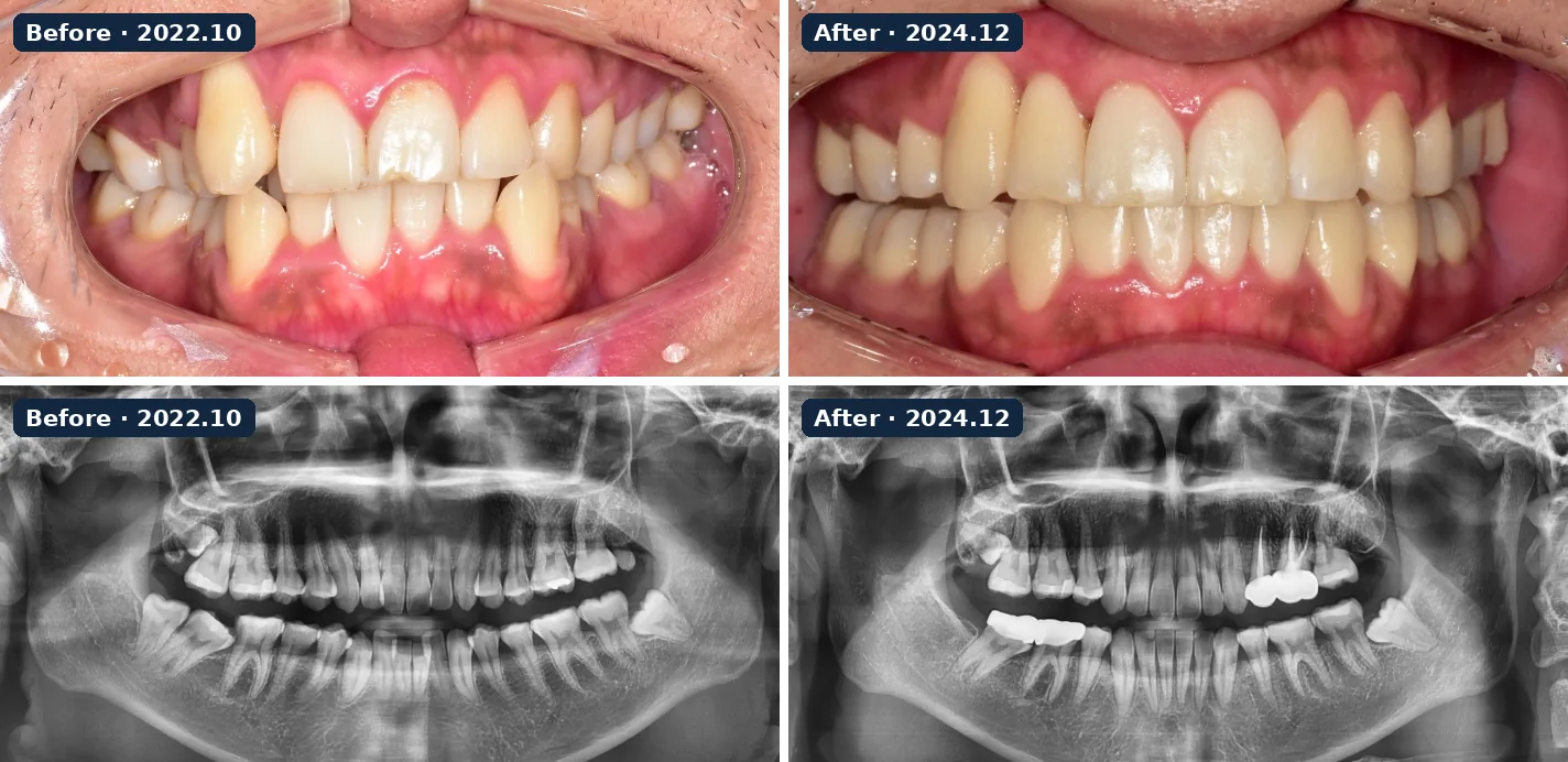
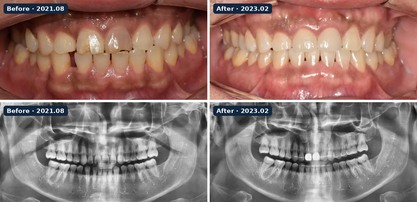
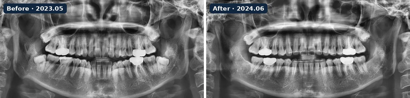
Ten orthodontic cases from the clinic’s Invisalign records, each showing the same patient before and after treatment with intraoral photos and panoramic X-rays (case 10: X-rays only). Published with the patients’ consent; before and after images were taken under the same conditions. Results differ for every patient. Orthodontic treatment can cause early discomfort, gum inflammation or other side effects, which your dentist explains and monitors. About Invisalign →
Orthodontic case 1 · 2023.06 → 2025.01 · intraoral photos and panoramic X-raysOrthodontic case 2 · 2021.01 → 2024.01 · intraoral photos and panoramic X-raysOrthodontic case 3 · 2023.05 → 2025.06 · intraoral photos and panoramic X-raysOrthodontic case 4 · 2018.04 → 2020.02 · intraoral photos and panoramic X-raysOrthodontic case 5 · 2021.11 → 2024.03 · intraoral photos and panoramic X-raysOrthodontic case 6 · 2022.06 → 2025.02 · intraoral photos and panoramic X-raysOrthodontic case 7 · 2022.01 → 2024.04 · intraoral photos and panoramic X-raysOrthodontic case 8 · 2022.10 → 2024.12 · intraoral photos and panoramic X-raysOrthodontic case 9 · 2021.08 → 2023.02 · intraoral photos and panoramic X-raysOrthodontic case 10 · 2023.05 → 2024.06 · panoramic X-rays
Clinical case studies
Six clinical cases, step by step
Published with the patient’s consent. Photos and X-rays are from the clinic’s records. Treatment plans and results differ for every patient; only an examination can show what is possible for you.
Full-arch implants
Full-arch implants after years of a loose lower denture
Treated by Dr. Hyeonjoon Park
Before
After
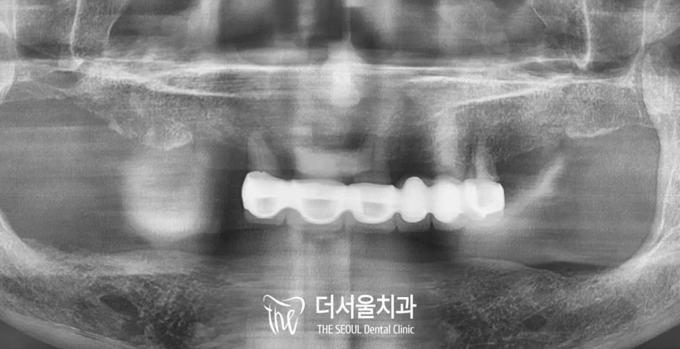
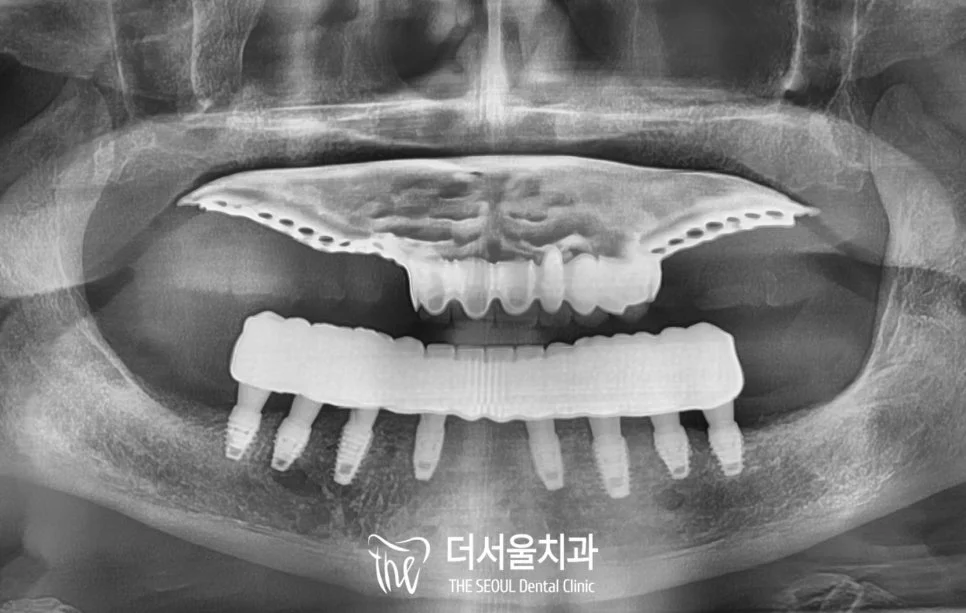
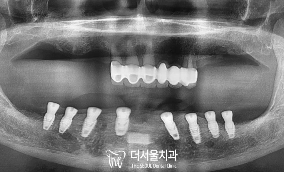
Panoramic X-rays before and after treatment
Before
After
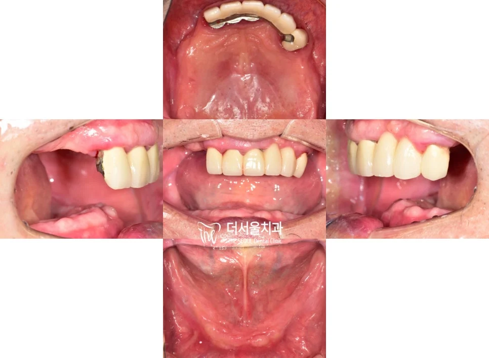
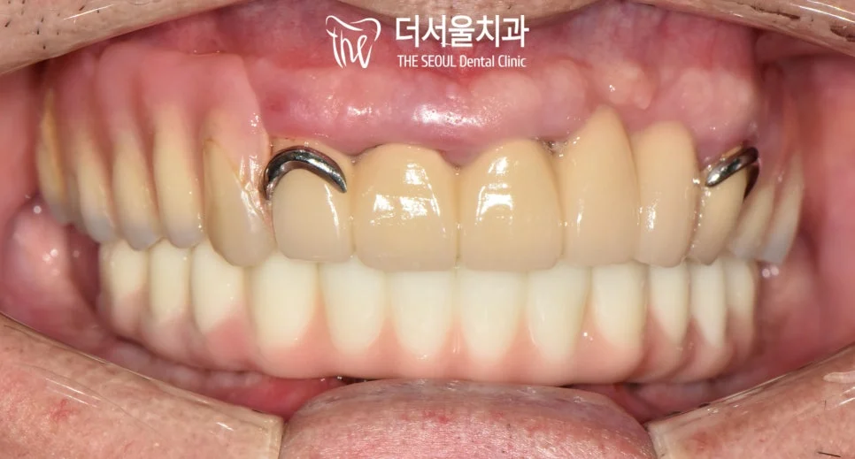
Intraoral photos before and after treatment
Why the patient came in
The patient had worn a lower denture for many years. It moved while eating and had to be removed and cleaned after every meal. The panoramic X-ray showed that long-term denture wear had left the lower jawbone low and thin.
What was done
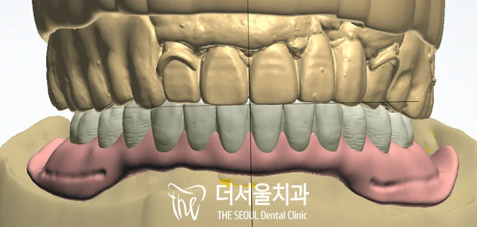
3D CT and an intraoral scan were used to plan implant positions, angles and lengths on the computer before surgery.
Eight implants were placed in the lower jaw, together with a bone graft.
Fixed temporary teeth were connected to the implants the day after surgery.
About three months later, custom abutments and the final full-arch prosthesis were fitted.
Digital design of the prosthesis from the scan dataX-ray after implant placement with temporary teeth
Published with the patient’s consent. Photos and X-rays are from the clinic’s records. Treatment plans and results differ for every patient; only an examination can show what is possible for you. Implant surgery can cause swelling, bruising or discomfort for some days, and implants need regular check-ups and careful cleaning to avoid peri-implantitis.
Implants
Redoing failed molar implant crowns and replacing a missing front tooth
Treated by Dr. Hyun Sung Park
Before
After
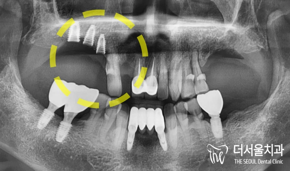
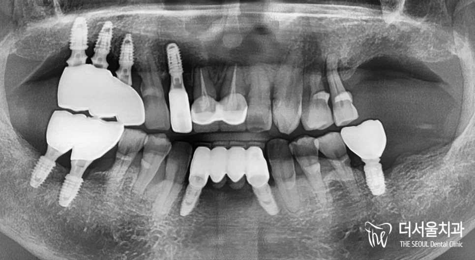
Panoramic X-rays before and after treatment
Why the patient came in
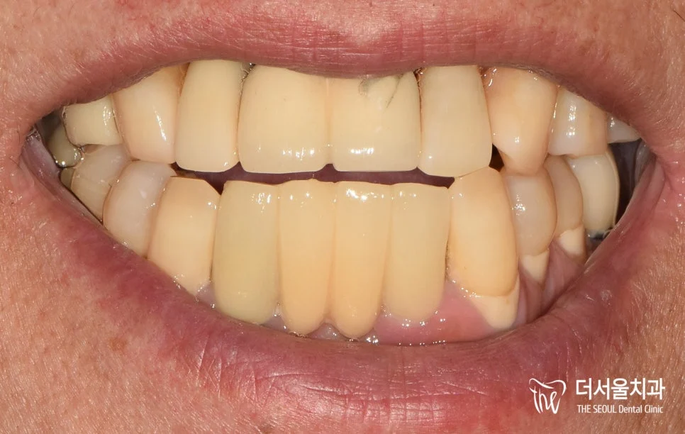
The patient came in after the restoration on three upper molar implants, placed at another clinic, had come off. The X-ray showed the three implant fixtures still in the bone, with gum and bone loss around them. An upper front tooth (#12) was also missing, and the front teeth were crowded.
What was done
CT and scan data were analysed on the computer to check bone density, crowding, mobility and undercuts before planning.
For the molars, new abutments and crowns were made for the existing implants.
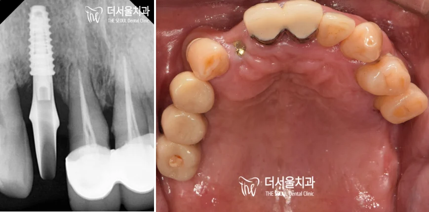
A narrow implant was placed for the missing front tooth (#12), and its crown was made after the implant had integrated with the bone.
The patient was advised to come for regular check-ups, because problems around implants often give no early pain.
Front-tooth implant: X-ray and the upper arch after treatmentAfter treatment
Published with the patient’s consent. Photos and X-rays are from the clinic’s records. Treatment plans and results differ for every patient; only an examination can show what is possible for you. Implant treatment can cause swelling or discomfort after surgery. Implants and their crowns need regular check-ups, because problems around an implant are often painless at first.
Orthodontics
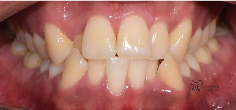
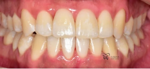
Crowded, protruding front teeth treated with Invisalign without extractions
Treated by Dr. Hyeonjoon Park
Before
After
Front view before and after treatment
Before
After
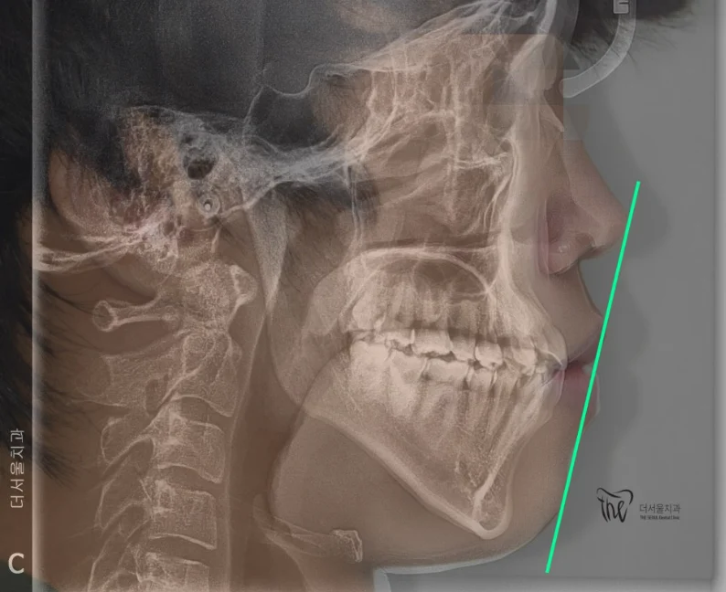
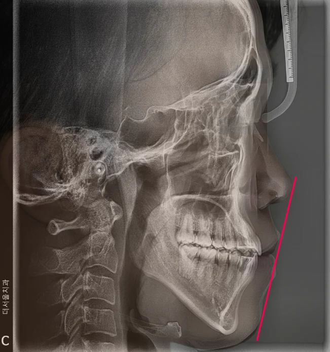
Lateral cephalometric X-rays before and after: lip position against the aesthetic line
Why the patient came in
The patient came in for a protruding mouth and crooked teeth. The front teeth were severely crowded and tilted inward, the dental arches were narrow, and the lips were strained when closed. Crowding like this also makes thorough brushing difficult.
What was done
The patient chose Invisalign clear aligners.
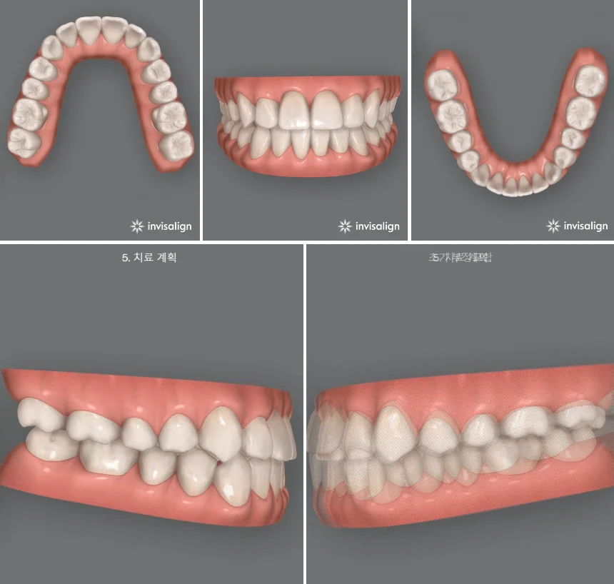
A ClinCheck simulation was used to plan the tooth movements and preview the expected result.
The arches were widened to create space for the crowded teeth, so no teeth were extracted.
At the end of treatment the teeth were evenly aligned and the lips sat behind the aesthetic line on the X-ray.
ClinCheck simulation of the planned result
Published with the patient’s consent. Photos and X-rays are from the clinic’s records. Treatment plans and results differ for every patient; only an examination can show what is possible for you. Orthodontic treatment can cause early discomfort, gum inflammation or root resorption. Retainers are needed afterwards to keep the result.
Cosmetic
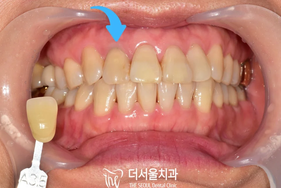
A darkened front tooth: root canal retreatment, internal whitening and a ceramic crown
Treated by Dr. Hyun Sung Park
Before
After
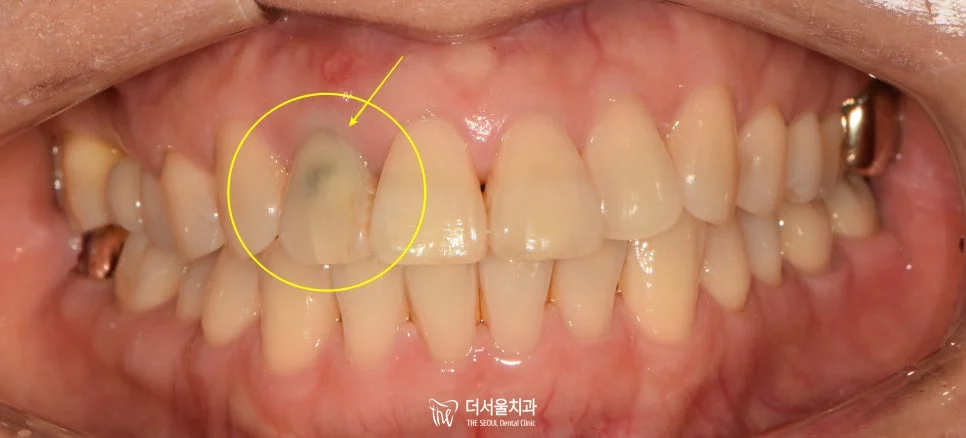
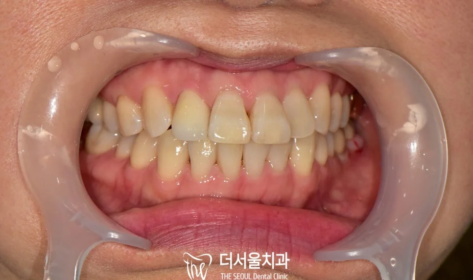
Front view before and after treatment
Why the patient came in
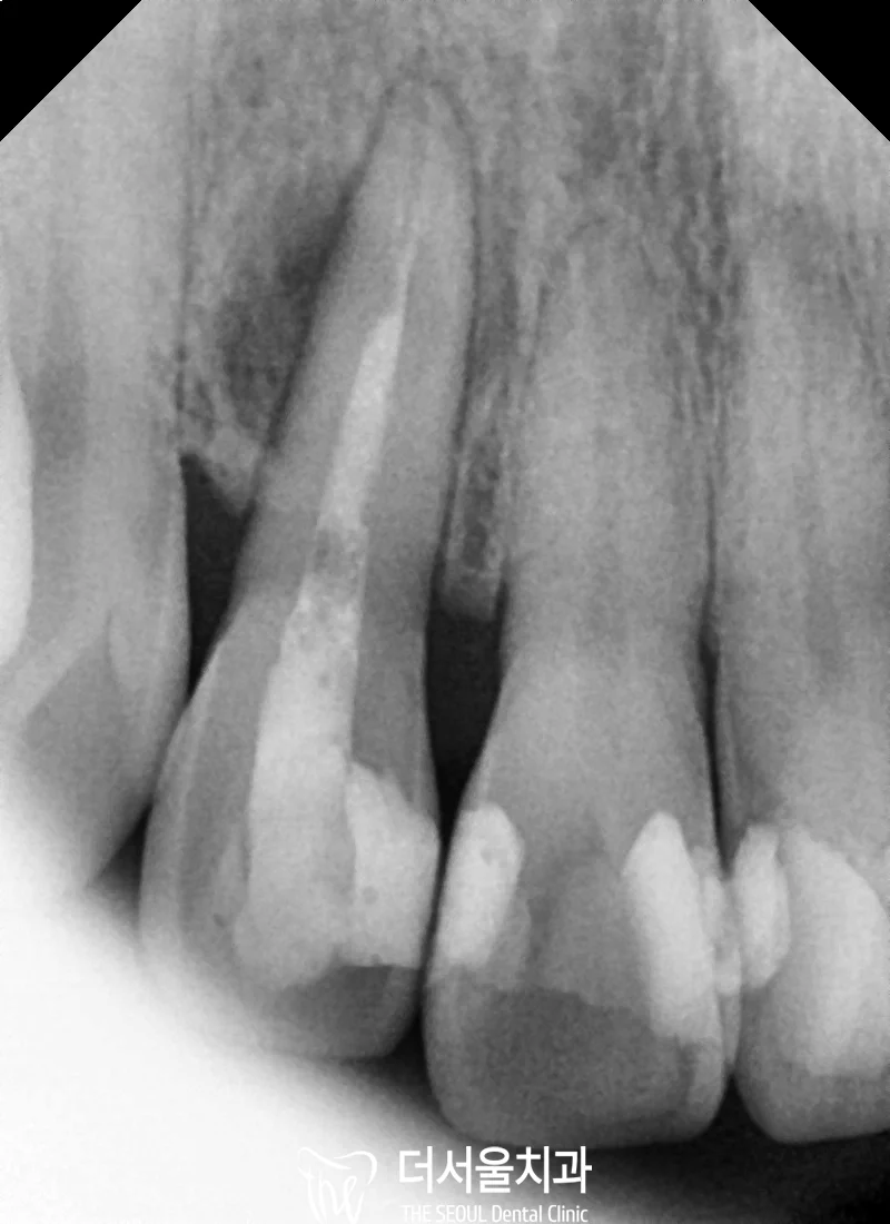
The patient came in because one upper front tooth had turned dark. The tooth had a root canal treatment from before, and the X-ray showed inflammation at the root tip.
What was done
The root canal treatment was redone. On opening the tooth, dark contaminated material was found inside the canal.
Part of the root filling was removed and a barrier of glass ionomer or resin was placed to keep the whitening agent inside the crown of the tooth.
A whitening agent was sealed inside the tooth and the colour was checked every 4 to 7 days.
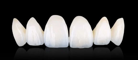
After whitening, the shade was matched and an all-ceramic crown was made so that no dark colour shows through.
X-ray before retreatmentShade check after internal whiteningCeramic crowns made to match the neighbouring teeth
Published with the patient’s consent. Photos and X-rays are from the clinic’s records. Treatment plans and results differ for every patient; only an examination can show what is possible for you. A tooth whitened from inside can darken again over time. Retreatment and crown preparation can cause temporary sensitivity or discomfort.
Cavity treatment
Deep decay under an old filling on a front tooth
Treated by Dr. Hyeonjoon Park
Before
After
Front view before and after treatment
Why the patient came in
An upper front tooth (#11) had extensive decay that had developed again around an old filling. The patient had throbbing pain even at rest. This was the last stage of a longer treatment for the same patient.
What was done
Cold and heat tests were used to check the nerve. Because of the pain at rest, root canal treatment was needed.
All decay was removed and the canal was cleaned and disinfected with the tooth kept dry.
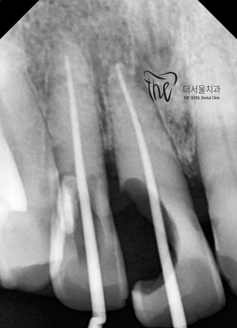
The canal was sealed with filling material to the root tip, checked on a high-resolution X-ray.
The tooth was restored with a zirconia crown.
X-ray of the sealed root canals
Published with the patient’s consent. Photos and X-rays are from the clinic’s records. Treatment plans and results differ for every patient; only an examination can show what is possible for you. Root canal treatment can cause tenderness for a few days, and a treated tooth can still fracture without a crown.
Root canal treatment
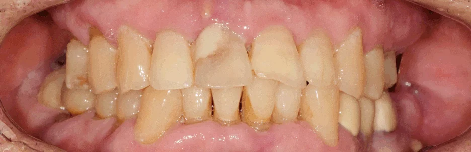
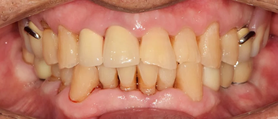
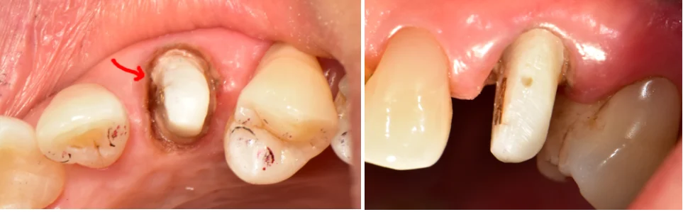
Why a crown matters after root canal treatment: saving a fractured canine
Treated by Dr. Hyeonjoon Park
Before
After
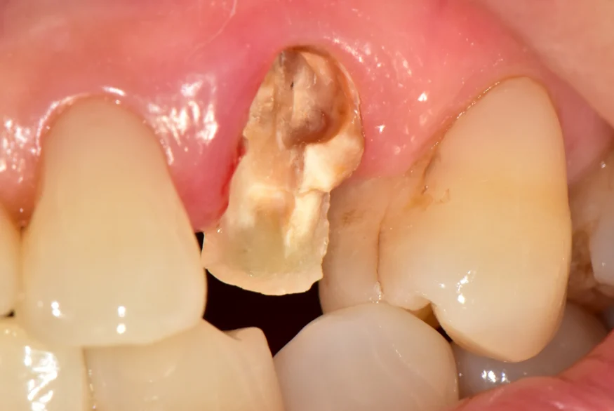
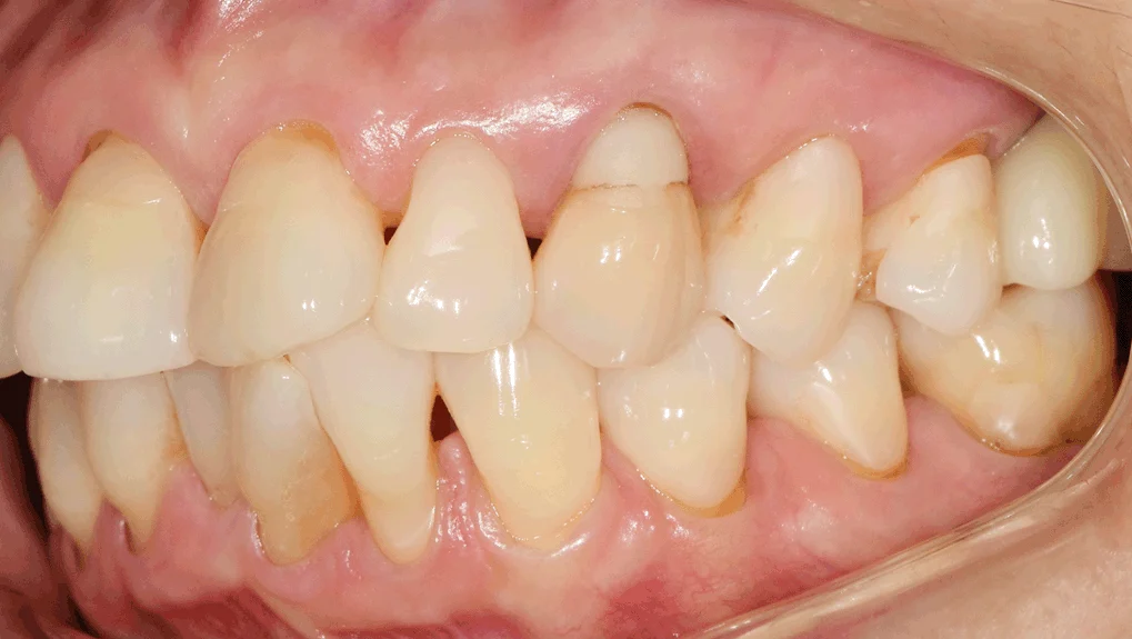
The fractured tooth and the final crown
Why the patient came in
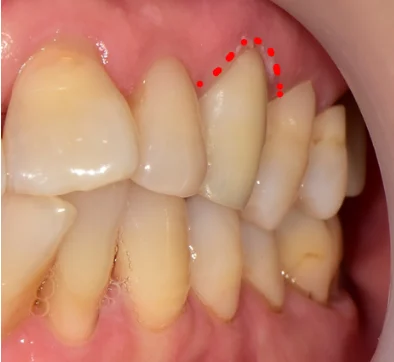
An upper canine (#23) had a root canal treatment done at another clinic but no crown, plus a large glass-ionomer filling from earlier gum-line wear. A crown was recommended at a check-up, but the patient chose to wait. Later the cheek side of the tooth broke off, leaving the tooth hollow.
What was done
The patient preferred to keep the tooth rather than have an implant.
The root canal treatment was redone.
Because very little tooth structure remained, a core build-up was made to give the crown enough support.
A crown was then fitted.
Before the fracture: root-canal-treated canine without a crownCore build-up before the crown
Published with the patient’s consent. Photos and X-rays are from the clinic’s records. Treatment plans and results differ for every patient; only an examination can show what is possible for you. A root-canal-treated tooth is more brittle, so a crown is usually advised. Retreatment can cause temporary tenderness.
Talk to us before you book
Send a short message in English. We will reply with what to bring and what to expect.